Four modules measure the radiograph you already take and return the angles, indices and suitability checks to the reporting surgeon. Assessment support — the clinician makes the diagnosis.
A pelvic or shoulder radiograph carries a dozen landmarks. Placing them manually takes minutes per study, and the result moves with the observer: the same film measured twice rarely gives the same number twice.
Inter-observer variability makes follow-up comparison unreliable, and the queue of pending reports grows. Nothing about that work is judgement — it is arithmetic on landmarks, and it can be handed to software that does it identically every time.
Each module ships with the measurement set below. Values shown are real outputs from the application.
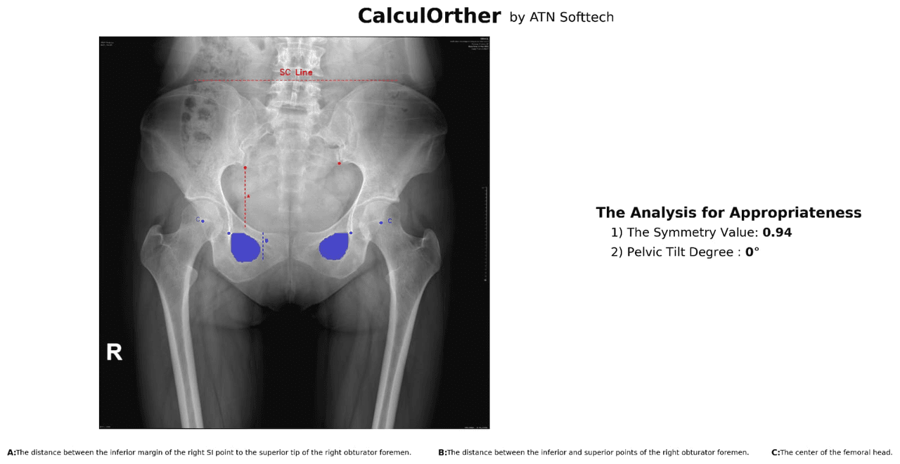
Before anything is measured, the film has to be worth measuring. PelSiAn checks obturator foramen symmetry and pelvic tilt and tells the technician whether the projection is suitable — the only automated suitability check the team has found in this field.
Suitability support for acquisition. Repeat exposures avoided rather than diagnoses made.
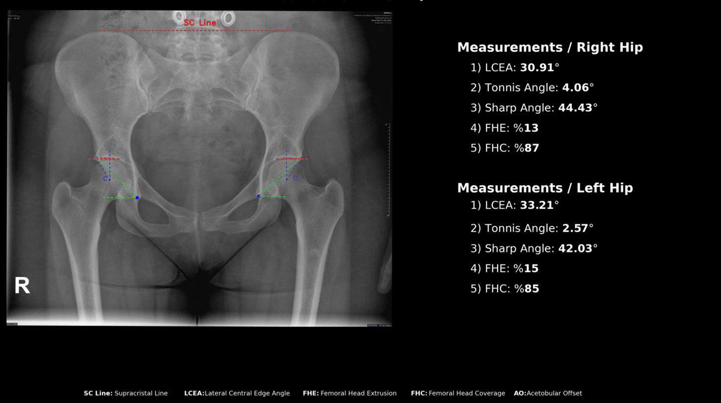
Femoroacetabular morphology on a standard AP pelvis: lateral centre-edge angle, Tönnis angle, Sharp angle, femoral head extrusion and acetabular coverage — both hips in one pass, with the landmarks left visible so the surgeon can correct them.
Morphometric assessment support; validated in AOTT 2026.
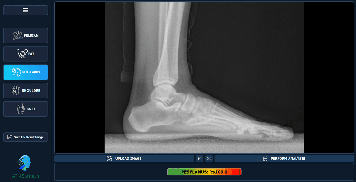
Weight-bearing lateral foot radiographs, measured for arch geometry, with the study list, overlay controls and export panel of the application around it. This is the module that shows how the software sits in a reporting session rather than in a demo.
Measurements are returned to the report; interpretation stays clinical.
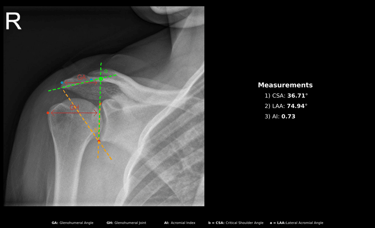
Critical Shoulder Angle, Lateral Acromion Angle and Acromial Index, drawn from the glenoid and acromion landmarks on a standard AP shoulder film. The three values that shoulder surgeons currently measure by hand, produced identically every time.
No competitor platform in the comparison below offers this shoulder set.
Yılmaz A., Selçuk T., Aksoy T., Atilla B. A quantitative analysis of symmetry on standard anteroposterior pelvic X-ray. Acta Orthopaedica et Traumatologica Turcica, 2025;59:122–128.
DOI 10.5152/j.aott.2025.24033
Academic studies on the module set were completed in December 2025. The other three publications cover the reliability of CalculOrther itself, the three shoulder angles and pes planus detection.
No commercialized domestic orthopedic X-ray analysis product exists in Türkiye; internationally, the closest platforms carry radiology suites rather than these specific orthopedic measurements.
| CAPABILITY | CALCULORTHER | Sectra | ImageBiopsy Lab | RAYLYTIC |
|---|---|---|---|---|
| Shoulder angle analysis (CSA · LAA · AI) | Yes | — | — | — |
| Pelvic radiograph suitability check | Yes | — | — | — |
| Pelvic tilt analysis | Yes | — | — | — |
| Hip / FAI morphometry | Yes | Partial | Yes | Yes |
| Pes planus / foot geometry | Yes | Partial | Partial | — |
| Works on plain radiographs only | Yes | Yes | Yes | Yes |
| Published validation in orthopedic journal | AOTT 2025 · 2026 | — | Yes | Yes |
| Domestic support and training (Türkiye) | Yes | — | — | — |
One annual institutional licence covers a single institution, unlimited users and all four modules. It runs against the existing PACS/RIS, and the licence includes staff training and support for the reporting workflow.
Pilot clinic · Niğde Ömer Halisdemir University Hospital Orthopedics & Traumatology